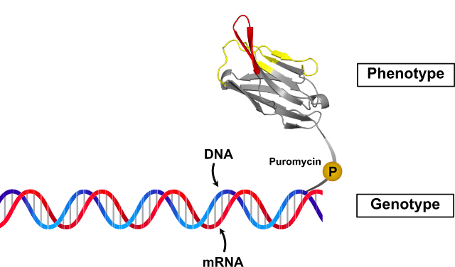
※Image of cDNA display molecule cDNA display is our protein display method which is genotype/phenotype linking systems. cDNA display has
high stability and it handles high diversity.
The Advantages of cDNA display
Restriction of Displayed protein
No restriction for proteins with cytotoxicity
cDNA display enables to display proteins which have cytotoxicity for cells and microbes.
Stability
Very high stability
Displayed protein is linking to cDNA display by linker.
Therefore, cDNA display is very stable in alkaline pH, high temperature, and RNase.
Diversity
Very huge diversity
Available to hundle approximately 10¹³⁻¹⁴
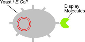
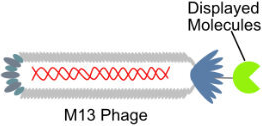
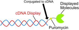
Comparison of cDNA display and other protein display method
Display method
Structure
Genotype
Diversity (/mL)
Yeast display
Plasmid DNA
10⁸
Phage display
ssDNA
10⁸
cDNA display
cDNA
10¹³⁻¹⁴
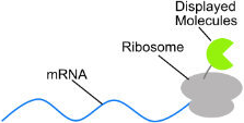
Ribosome display
mRNA
10¹³
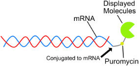
mRNA display
mRNA
10¹³
EME’s linker technology
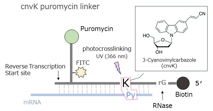
In the novel cnvK-based puromycin linker cDNA display method, our proprietary cnvK linker is used as the puromycin linker, which is the key technology for genotype-phenotype mapping. UV irradiation of mRNA and cnvK linker hybridized at a specific site rapidly link the mRNA and cnvK linker by photocrosslinking, forming an mRNA-cnvK linker complex. This has enabled a significant reduction in the time required to prepare of cDNA display molecules.
The advantages of using the cnvK linker are as follows; ● Linking mRNA and puromycin linker by photocross-linking instead of enzymatically => shorter reaction time(about 1 hour => a few minutes) ● Reduction of contamination such as impurities from the enzyme solution is expected to improve the reaction efficiency of the cDNA display molecule. ● Can be used for both in vitro selection experiments and evaluation of candidate clone binding
The method of genotype-phenotype linking strategy with puromycin linker
After the preparation of the mRNA-cnvK linker, the mRNA-cnvK linker is added to cell free translation
reaction mixture. Ribosome starts translation from 5’ ends side of mRNA, and polypeptides are synthesized.
Because ribosome doesn’t translate the sequence of cnvK linker, ribosome stops translation when ribosome
reaches the sequence of cnvK linker. At the same time, puromycin (aminoacyl tRNA analog) in the cnvK linker
is integrated into ribosome. When the cnvK linker is integrated into ribosome, the peptide transfer reaction
is occurred. Because of the peptide transfer reaction, the C-termini of synthesized polypeptide chain and
puromycin linker is connected with covalent bond. Therefore, this synthesized compound is mRNA-cnvK linker
polypeptide. In EME, we use our unique linker “photo-cross linkage puromycin linker (cnvK linker)” and “cDNA display
synthesizer.” These technologies allow the auto preparation of large diversity (10¹³⁻¹⁴) of cDNA library.
EME’s original cnvK linker
By applying EME’s proprietary cnvK linker to screening technology, clones can be screened from a vast library in a short period of time. If you are interested in cnvK linker, please contact us using the contact form below. Contact us from here